Human Leg Bone Diagram / WY_4907 Fibula Neck Diagram Wiring Diagram : These bones are arranged into two major divisions:
byAdmin-
0
Human Leg Bone Diagram / WY_4907 Fibula Neck Diagram Wiring Diagram : These bones are arranged into two major divisions:. There also are bands of fibrous connective tissue—the ligaments and the tendons—in intimate relationship with the parts of the skeleton. Distal end of right humerus. The human leg consists of 8 bones, 4 per leg. Human skeleton long bones of arms and legs britannica. He legs provide support for the body and power much of its movement.
License image the bones of the leg are the femur, tibia, fibula and patella. Leg bone anatomy diagram diagram of human leg human anatomy. The femur, or thighbone, is the longest and largest bone within the human physique. Your leg bones are the longest and strongest bones in your body. There also are bands of fibrous connective tissue—the ligaments and the tendons—in intimate relationship with the parts of the skeleton.
Bones of foot, labeled diagram. poster | Zazzle.com from rlv.zcache.com It is the most complete reference of human anatomy available on web, ipad, iphone and android devices. The human leg consists of 8 bones, 4 per leg. This framework consists of many individual bones and cartilages. He leg's main function in the human is for locomotion and support of the rest of the body. High resolution textures and displacement included. Alibaba.com offers 851 human leg bone products. Related posts of diagram of leg bones nasal bone anatomy x ray. Let's assume this figure is standing with feet vertically aligned with the hip joints.
High quality realistic skeleton legs.
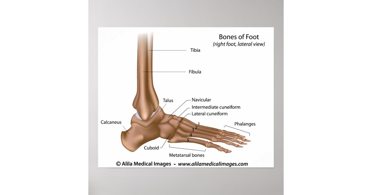
He leg's main function in the human is for locomotion and support of the rest of the body. Distal end of right humerus. Download this free vector about diagram showing the hip bone treatment, and discover more than 11 million professional graphic resources on freepik. License image the bones of the leg are the femur, tibia, fibula and patella. Vector illustration anatomy of human legs and diagram of human bones isolated on white background. Human anatomy for muscle, reproductive, and skeleton. The foot bones shown in this diagram are the talus, navicular, cuneiform, cuboid, metatarsals and calcaneus. The foot bones shown in this diagram are the talus, navicular, cuneiform, cuboid, metatarsals and calcaneus. The foot bones shown in this diagram are the talus, navicular, cuneiform, cuboid, metatarsals and calcaneus. He leg's main function in the human is for locomotion and support of use the leg bones diagrams to learn the names of the leg bones and leg anatomy. When you stand or walk, all the weight of your upper body rests on them. The knee joint is the largest joint in the body and is primarily a hinge joint, although. There also are bands of fibrous connective tissue—the ligaments and the tendons—in intimate relationship with the parts of the skeleton.
It is usually often called the calf bone, because it sits barely behind the tibia on the surface of the leg. He legs provide support for the body and power much of its movement. Human muscle system the muscles of the. Includes leg (femur, tibia, patella, and fibula) and foot (tarsals and digits) bones. Human anatomy diagrams show internal organs, cells, systems, conditions, symptoms and sickness information and/or tips for healthy living.
16 best Bones in the Leg images on Pinterest | Human body ... from i.pinimg.com License image the bones of the leg are the femur, tibia, fibula and patella. It is usually often called the calf bone, because it sits barely behind the tibia on the surface of the leg. The human leg consists of 8 bones, 4 per leg. The femur, or thighbone, is the longest and largest bone within the human physique. Human muscle system the muscles of the. Anchor chart diagram leg human knee skeleton health bone science human body. Download this free vector about diagram showing the hip bone treatment, and discover more than 11 million professional graphic resources on freepik. Disposition of rotator cuff muscles diagram.
He leg's main function in the human is for locomotion and support of the rest of the body.
Leg bone anatomy diagram diagram of human leg human anatomy. The knee joint is the largest joint in the body and is primarily a hinge joint. The humerus is the (upper) arm bone. It is the most complete reference of human anatomy available on web, ipad, iphone and android devices. Joints of hand anterior view, lateral view, right hand. Related posts of diagram of leg bones nasal bone anatomy x ray. The foot bones shown in this diagram are the talus, navicular, cuneiform, cuboid, metatarsals and calcaneus. Vector illustration anatomy of human legs and diagram of human bones isolated on white background. As these muscles contract and relax they move skeletal bones to create movement of the body. He leg's main function in the human is for locomotion and support of use the leg bones diagrams to learn the names of the leg bones and leg anatomy. Separate files for each identical bones. Human muscle system the muscles of the. The human leg consists of 8 bones, 4 per leg.
Foot and ankle diagram anatomy. File is ready to render. Leg bone anatomy diagram diagram of human leg human anatomy. Anatomy diagram of human leg bone structure. When your muscles contract, they pull the bone they're.
Broken Leg, Pictures, Causes, Treatment, Surgery ... from images.emedicinehealth.com Synovial joint capsule bones chart. Vector illustration anatomy of human legs and diagram of human bones isolated on white background. The bones of your leg have roughened patches on their surfaces where muscles are attached. As these muscles contract and relax they move skeletal bones to create movement of the body. When your muscles contract, they pull the bone they're. They are named by region formed by the left and right hip bones, the pelvic girdle connects the lower limb (leg) bones to the axial skeleton. He leg's main function in the human is for locomotion and support of the rest of the body. Joints of hand anterior view, lateral view, right hand.
Let's assume this figure is standing with feet vertically aligned with the hip joints.
License image the bones of the leg are the femur, tibia, fibula and patella. Your leg bones are the longest and strongest bones in your body. The foot bones shown in this diagram are the talus, navicular, cuneiform, cuboid, metatarsals and calcaneus. Lower bones limbs limb leg diagram muscle foot template anatomy blank human skeleton coloring sketch function th. He legs provide support for the body and power much of its movement. The human leg, in the general word sense, is the entire lower limb of the human body, including the foot, thigh and even the hip or gluteal region. Download this free vector about diagram showing the hip bone treatment, and discover more than 11 million professional graphic resources on freepik. The foot bones shown in this diagram are the talus, navicular, cuneiform, cuboid, metatarsals and calcaneus. 10 amazing facts about the human body you didn't know before 2021 | human body mystery. There also are bands of fibrous connective tissue—the ligaments and the tendons—in intimate relationship with the parts of the skeleton. Synovial joint capsule bones chart. Human muscle system the muscles of the. They are named by region formed by the left and right hip bones, the pelvic girdle connects the lower limb (leg) bones to the axial skeleton.
Separate files for each identical bones leg bone diagram. Let's assume this figure is standing with feet vertically aligned with the hip joints.